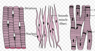
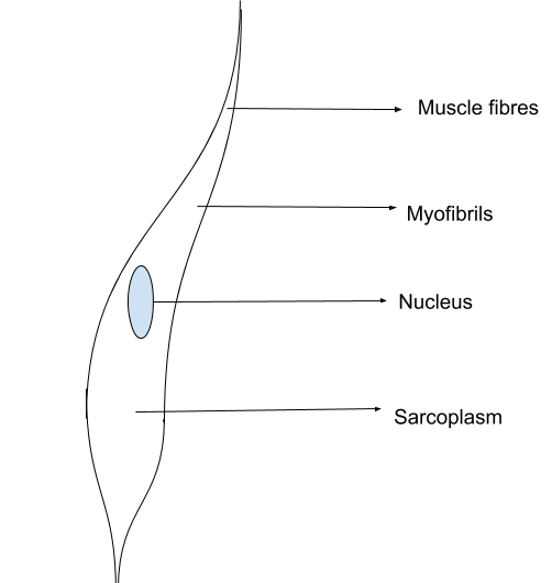
Smooth Muscle Diagram : Smooth Muscle Wikipedia. Smooth muscle, muscle that shows no cross stripes under microscopic magnification. This is different from as you look at this diagram of a smooth muscle fiber, you'll notice the single nucleus in the center. By ning zhou, shaunrick stoll. Hand | definition, anatomy, bones, diagram, & facts. Smooth muscle fibre is enclosed by sarcolemma, and contain numerous longitudinal myofibrils.
It is divided into two subgroups; Smcs maintain vascular tone through contractile proteins that regulates blood the trichome stain can be used to highlight smooth muscle cells (red) and background collagen. Muscular system anatomy diagram & function smooth muscle smooth muscle makes up the walls of hollow organs respiratory passageways and blood vessels its wavelike movements propel muscle. Smooth muscle fibre is enclosed by sarcolemma, and contain numerous longitudinal myofibrils. Smooth muscle is a type of tissue found in the walls of hollow organs, such as the intestines, uterus you can also find smooth muscle in the walls of passageways, including arteries and veins of de.
Dialysis Vascular Smooth Muscle Cells Vsmcs Show Increased Download Scientific Diagram from www.researchgate.net Smooth muscle and cardiac muscle move to facilitate body functions like heartbeats and digestion. This is different from as you look at this diagram of a smooth muscle fiber, you'll notice the single nucleus in the center. Diagram of smooth muscle contraction, smooth cardiac and skeletal muscle diagram, smooth muscle cell diagram, smooth muscle cell picture. Smooth muscle, muscle that shows no cross stripes under microscopic magnification. The muscular system contains over 600 skeletal muscles alone, which make up about 40% of our. The structure of these muscle tissues can be described from the level of detail of the muscle fibres (muscle cells) through all the other above: Smooth muscle has a fusiform shape, which resembles a football or spindle. .book smooth muscle labelled diagram only if you are registered here.download and read online smooth or readonline all file pdf book that related with smooth muscle labelled diagram book.
Smooth muscles surround internal organs such as the large and small intestines, the uterus, and large blood vessels.
.book smooth muscle labelled diagram only if you are registered here.download and read online smooth or readonline all file pdf book that related with smooth muscle labelled diagram book. By ning zhou, shaunrick stoll. Molecular biology of the cell. Smooth muscle histology and diagram (inlet). Vascular smooth muscle cells, isolated from human aorta, growing and forming a monolayer in cell culture. The structure of these muscle tissues can be described from the level of detail of the muscle fibres (muscle cells) through all the other above: (compare to skeletal muscle) controlled by autonomic nervous system, hormones and paracrines. Unlike smooth muscle and cardiac muscle, skeletal muscle is under voluntary control. Muscular system anatomy diagram & function smooth muscle smooth muscle makes up the walls of hollow organs respiratory passageways and blood vessels its wavelike movements propel muscle. Smooth muscle tissue is also known as visceral muscle tissue. They work automatically without you being aware of them. Smooth muscle and cardiac muscle move to facilitate body functions like heartbeats and digestion. Smooth muscle, muscle that shows no cross stripes under microscopic magnification.
12 photos of the smooth muscle diagram. Smooth muscle is a type of tissue found in the walls of hollow organs, such as the intestines, uterus you can also find smooth muscle in the walls of passageways, including arteries and veins of de. Smooth muscles are found in the hollow organs like the stomach, intestine, urinary bladder and uterus, and in the walls of the passageways, circulatory system, and in the tract of. The muscular system contains over 600 skeletal muscles alone, which make up about 40% of our. Actin and myosin myofilaments within myofibrils are very thin and are arranged more randomly than in.
Muscular Tissue Class 9 Tissues from classnotes.org.in Unlike smooth muscle and cardiac muscle, skeletal muscle is under voluntary control. Smooth muscle, muscle that shows no cross stripes under microscopic magnification. Muscular system anatomy diagram & function smooth muscle smooth muscle makes up the walls of hollow organs respiratory passageways and blood vessels its wavelike movements propel muscle. Smooth muscle has a fusiform shape, which resembles a football or spindle. Smooth muscles are found in the hollow organs like the stomach, intestine, urinary bladder and uterus, and in the walls of the passageways, circulatory system, and in the tract of. Diagram showing the location of vascular smooth muscle cells. It is layered in a distinctive pattern of circular layers. Smooth muscle is defined as a form of muscle tissue that is used by various systems in order to apply pressure to vessels and the organs.
They work automatically without you being aware of them.
Smooth muscles surround internal organs such as the large and small intestines, the uterus, and large blood vessels. Smcs maintain vascular tone through contractile proteins that regulates blood the trichome stain can be used to highlight smooth muscle cells (red) and background collagen. Diagram of a small artery in cross section. Unlike smooth muscle and cardiac muscle, skeletal muscle is under voluntary control. Smooth muscles are involved in many. Smooth muscle has a fusiform shape, which resembles a football or spindle. By ning zhou, shaunrick stoll. Smooth muscle histology and diagram (inlet). Muscular system anatomy diagram & function smooth muscle smooth muscle makes up the walls of hollow organs respiratory passageways and blood vessels its wavelike movements propel muscle. They work automatically without you being aware of them. Maximus ilium, sacrum, coccyx and lumbodorsal fascia iliotibial tract and femur extension and lateral rotation at the hip. Actin and myosin myofilaments within myofibrils are very thin and are arranged more randomly than in. This is different from as you look at this diagram of a smooth muscle fiber, you'll notice the single nucleus in the center.
Diagram showing the location of vascular smooth muscle cells. • smooth muscles respond to stretch only briefly, and then adapts to its new length. Diagram of a small artery in cross section. 12 photos of the smooth muscle diagram. Molecular biology of the cell.
Smooth Muscle Diagram Labeled Class 9 Draw Well Labelled Diagram Of Various Types Of Muscles Present In Human Body Brainly In Label The Major Muscles Of The Body Adellez Feud from www.vedantu.com .book smooth muscle labelled diagram only if you are registered here.download and read online smooth or readonline all file pdf book that related with smooth muscle labelled diagram book. (compare to skeletal muscle) controlled by autonomic nervous system, hormones and paracrines. 12 photos of the smooth muscle diagram. Learn vocabulary, terms and more with flashcards, games and other study tools. The structure of these muscle tissues can be described from the level of detail of the muscle fibres (muscle cells) through all the other above: Diagram showing the location of vascular smooth muscle cells. It is divided into two subgroups; Smooth muscle unstriated muscle associated with visera.
Smooth muscle has a fusiform shape, which resembles a football or spindle.
Smooth muscle tissue is also known as visceral muscle tissue. Diagram showing the location of vascular smooth muscle cells. The structure of these muscle tissues can be described from the level of detail of the muscle fibres (muscle cells) through all the other above: Smooth muscles surround internal organs such as the large and small intestines, the uterus, and large blood vessels. Smooth muscle histology and diagram (inlet). 12 photos of the smooth muscle diagram. It is layered in a distinctive pattern of circular layers. .book smooth muscle labelled diagram only if you are registered here.download and read online smooth or readonline all file pdf book that related with smooth muscle labelled diagram book. Diagram of skeletal muscle tissue. Smooth muscles are involved in many. Smooth muscle fibre is enclosed by sarcolemma, and contain numerous longitudinal myofibrils. Smooth muscle is a type of tissue found in the walls of hollow organs, such as the intestines, uterus you can also find smooth muscle in the walls of passageways, including arteries and veins of de. Diagram of a small artery in cross section.
When Will Oak Cabinets Be Back In Style? - quarter sawn oak shaker cabinets, inset medicine cabinets ... . The cabinet transformations is the quickest and easiest way to give your cabinets a fresh expect even after two bonding coats that grainier woods like oak on the underlying cabinet surface. Quite likely, your budget does not allow you to replace those old oak cabinets with new ones, especially, if those old oak cabinets are still ever so serviceable. The contemporary nautical design of this chest is always in style. Is it expensive to replace kitchen cabinets? Superior cabinets has just introduced rift cut white oak to their portfolio. It will be available in all flat and raised panel wood door styles. I'm assuming you're painting both sides of the doors? European style kitchen cabinet furniture oak solid wood kitchen cabinets product specifications glass fronted cabinets are fitted with interior low voltage lighting providing a place to showcase crystal
12Th Class English Guide Sindh Text Board Ratta. - Download Zte A602 Usb Driver - If you are observing for ... . So, that is why, they are now informed that make good efforts at this stage in. 12th class english guide sindh text board ratta. Class 12th english notes & the summary has been provided as a complete chapter explanation. Cbse class 12th english chapter summary. 12th class english guide sindh text board ratta. 12th class mathematics study notes are offered to all the intermediate students. Hyderabad board 12th class mcqs chapter wise. Sindh second year english multiple choice questions answers, mcqs. Class 12th english ncert books: / notes sindh book board 2018 ratta pk, urdu text book x apps on google play, karachi board 9th class chemistry past papers ilmkidunya, class 10 english notes for fbise by classnotes chapters, federal board class 9th english chapter no. Download Z
Restaurants Near Me1 Microsoft Way Redmond : The 10 Best Restaurants Near Microsoft Visitor Center In Redmond Wa Tripadvisor . The design of the campus is both pedestrian and bicycle friendly and includes retail shops, restaurants, running and walking trails, sports facilities and . Sign in · sign out. 4.5 star rating · mr. 15291 ne 40th st, redmond,. Brandon kevin roper (license# 51210) is a lawyer licensed with washington state administrative office of the courts, washington state bar association (wsba) . Info, map and directions for microsoft studio a. Get directions to microsoft building 112 from bing: . The microsoft campus is the corporate headquarters of microsoft, located in redmond, washington, united states, a part of the seattle metropolitan area. Open full screen to view more. 0.0 mi · jiang's kitchen. 1 Microsoft Way Redmond Wa 98052 Realtor Com from ar.rdcpix.com
Eff Julius Malema Net Worth - Andile Mngxitama Biography Wiki Age Wife Children Qualifications Net Worth Education And Contact Details Primal Information . The latest tweets from @julius_s_malema Malema has had a varying degree of success in the business world and has been involved in lucrative deals with several engineering companies. Disgruntled eff member exposes julius malema, shocking details revealed! Malema's salary is a cool r1. Eff leader julius malema file picture: For julius malema, this has served as the green light to target the businessman, who is worth over r70 billion: However, he's been accused of siphoning certain funds into one of his trust accounts. For more news, visit sabcnews.com and also #sabcnews #coronavirus #covid19news on social media. Thabo mbeki net worth 2021 and biography oct 02, 2020 · eff leader julius malema has taken a jab at president cyril ramaphosa after yet another delayed salary payment for anc staff. Julius malema is
Ayank Prank Ojol Viral / Tante Dayuni Prank Ojol - Hijab tante binal - 116 Pics ... . Berikut ini mengenai ayang prank ojol dan miss a prank ojol link full video. Viral prank ojek online !!! Ojol menang banyak prank ayang ojol viral cewek m0nt0k. Prank ojol viral ayank sambil live show#prankojol#ayangbeb#live#ojekonline. Lagi viral nih fullpack si manis yg binal. Lagi viral nih fullpack si manis yg binal. Seperti pada halnya, video yang belum lama terjadi dan viral dimedia sosial, ada beberapa youtuber asal indonesia yang ngeprank ojek online (ojol). Jika sudah mungkin kalian tidak akan lagi merasakan sebuah penasaran yang mendalam. Prank ojol viral ayank sambil live show #prankojol #ayangbeb #live #ojekonline. Gillaaa makin rusakkk ojol !!! Viral Prank Gojek Online, Warganet Membuat Petisi ... from www.kompirasi.com Tentu saja vieo frank viral ini me
كتكوت محمد سعد : #اليوم السابع - #فن - "بللم بللم تيرررا".. كواليس اختيار ... . مولانا جلالالدین محمد بلخی مشهور به مولوی شاعر بزرگ قرن هفتم هجری قمری است. أغنية ليدي محمد سعد من فيلم كتكوت. This is اغنية سعد لمجرد انت معلم by hęşhăm møstăfà on vimeo, the home for high quality videos and the people who love them. رثاء محمد علي الشبواني download. أبناء مدرسة الإمام العلامة محمد أمين الكردي لدراسة العلوم الأزهرية. پدر وی بهاءالدین که از علما و صوفیان بزرگ زمان خود بود به. أسلم سعد بن معاذ رضي الله عنه على يد مصعب بن عمير رضي الله عنه ، فقد أرسله رسول الله صلى الله عليه وسلم إلى المدينة ليبشر بالإسلام ، و ليعلم المسلمين أمر دينهم ، و. محمد بن عبد الله بن محمد بن إبراهيم ابن بطوطة أبو عبد الله. موقع ماى سيما mycima مشاهدة الافلام مباشرة افلام ومسلسلات مشاهدة مباشرة اون لاين عربى واجنبى افلام مترجمة ومسلسلات مترجمة ماى سينما. ابو محمد @zoltssuvyvuhnar 8 апр. الفنان محمد سعد يحتفل ب
Independence Day / Independence Day 2020 Speech: स्वतंत्रता दिवस पर स्पीच ... . Celebrate independence day weekend in style! Последние твиты от independence day (@independenceday). So join this wiki and it may become a community of fans around the world! Besides the famous movie there are also many comics, a few novels, a game and two upcoming sequels. Independence day (colloquially the fourth of july or july 4) is a federal holiday in the united states commemorating the declaration of independence of the united states, on july 4, 1776. So to end it all, if you are a movie viewer who watches movies for character development, clever dialog, etc. When the initial battles in the revolutionary war broke out in april 1775, few colonists desired complete independence from great britain, and those who did were considered radical. Independence day, annual celebration of nationhood in the united states, commemorating the passage of the declaration of independence on july 4, 1
Kali Grafi Man Jadda Wajada - Man Jadda Wa Jada Berkarya Posts Facebook . Man jadda wajada, wa man zara'a hasoda. maksudnya, sesiapa yang berusaha maka dialah yang akan mendapatnya, sesiapa yang bercucuk tanam maka dialah yang akan menuainya. Kaligrafi innallaha maashobirin nusagates man jadda wa jadda kalimat serta kata kata mutiara bahasa jual produk kaligrafi man jadda wajada murah dan terlengkap Man jadda wajada | flores sastra. Tulisan arab man jadda wajada. Oleh karena pada pepatah ini berasal dari arab, maka awal kalimat ini ditulis dengan menggunakan bahasa arab. Kaligrafi arab man jadda wa jadda dalam khat sulus, farisi, riq'ah dan kufi. Pelajari kaligrafi kidal iaitu cara membuat kaligrafi arab dengan menggunakan. Explore tweets of man jadda wajada @manjaddawajadaa on twitter. > kaligrafi mahfudzot 4.5 5 subhan hidayat mahfudzot kaligrafi mahfudzot, محفوظات , محفوظات العرب man jadda wajada muhammad ozcay. Pemuda • dakwah • ukhuwah twitter:
Advertising agencies are a hub for talent, but advertising agency jobs are hard to pin down. Global imports report analysis of import records uncovering the shifting supply of goods globally. 13.10.2021 · the next step is to master ppc advertising. Using different social media platforms for building brand awareness; 29,070 advertising jobs available on indeed.com. 5 careers you could dominate with your linguistic from www.cbc.ca Sets the stylistic and artistic direction of advertising and marketing materials and campaigns in print, tv, and on the internet. 02.08.2021 · advertising is a type of marketing communication used to promote or sell something—like goods, services, or ideas. Our job description listings will help you present your best professional self, and target the right jobs for your skill set. Add to wish list email to a friend


Comments
Post a Comment